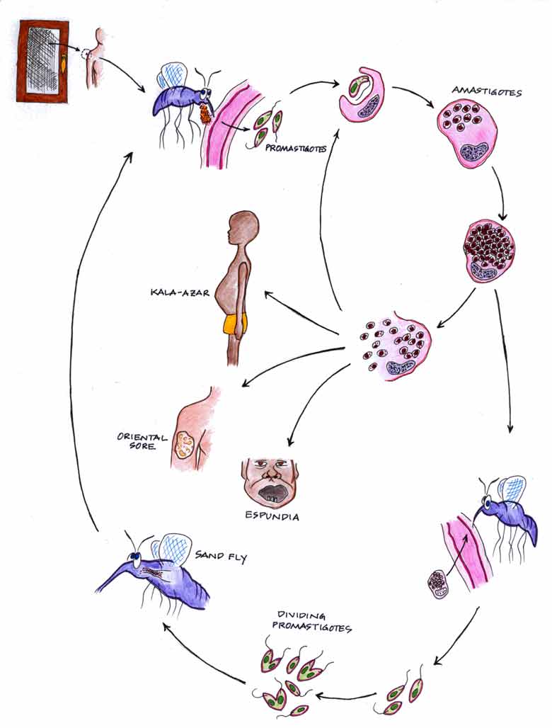
Leishmania spp. is the causative agent of a number of diseases in diverse locations in the world, roughly referred to as leishmaniasis. Though as many as 20 different species are recognized by some parasitologists, I will limit this discussion to five, Leishmania donovani, L. tropica, L. major, L. braziliensis and L. mexicana. L. donovani, L. tropica and L. major are transmitted by sand flies of the genus Phlebotomus, while L. braziliensis and L. mexicana are transmitted by the sand fly genus Lutzomyia. Sandflies are small bloodsuckers capable of passing through ordinary screen mesh in windows and doors. They live below 2000 feet above sea level, usually on plains. These "no-see-ums" suck blood and ingest amastigote forms from infected hosts. The amastigotes transform into flagellated promastigotes which clog the esophagus of the fly. Feeding flies, regurgitate promastigotes on the skin of the victim where they may enter the bite wound. Amastigotes of the different species are indistinguishable and represent one of the smallest eukaryotic cell known. Speciation is dependent on the geography of the infection as well as symptoms and disease states of the host. Visceral leishmaniasis is caused by L. donovani and affects mostly the spleen and liver. It is known in India as Dum-Dum fever or Kala-azar though it is much more widely distributed. It causes pronounced hepatosplenomegaly. Cutaneous leishmaniasis is caused by L. tropica and L. major causing skin lesions known as oriental sore, Baghdad boil, Delhi boil and other common names. The latter two leishmania are biochemically distinct, cause slightly different skin lesions, and do not overlap geographically. L. braziliensis is the cause of mucocutaneous leishmaniasis ranging from Mexico to mid-South America. Mucocutaneous leishmaniasis causes skin lesions which may be open and oozing, potentially spreading amastigotes to other parts of the body or other individuals. In some parts of South America, the disease known as espundia or uta refers to secondary lesions occuring in the nasal passageways or mouth, causing disfiguring sores and gaping holes. Sometimes the nasal septum, soft palate and other nasal/oral tissues are eaten away. Secondary infections may occur coincidentally with primary infections or many years after the primary infection has healed. L. mexicana
is found in Mexico, northern Central America, and Texas where it commonly causes a cutaneous lesion, though nasal/pharyngeal and visceral infections have been seen. Lumbermen, referred to as chicleros in Latin America, often display the badge of their occupation as an eroded ear.....chiclero ulcer.....due to an infection which has disintigrated some of the poorly-vascularized and therefore less immunocompetent ear cartilage.